Thursday, December 1, 2011

Cat Digestive System

Liver:
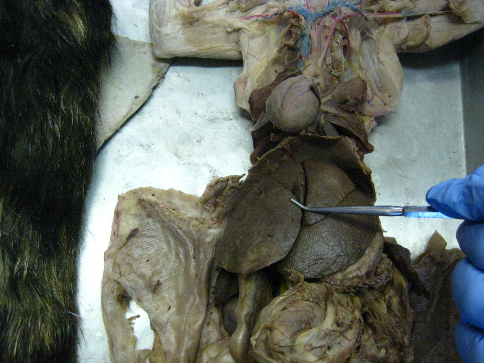
Cat's liver has multiple lobes, whereas a human liver has only 2 lobes: 1 right lobe and 1left lobe.


Gallbladder:


Common Bile Duct (of Gallbladder):


Falciform ligament:
Falciform ligament attaches the Liver to the anterior body wall.

Stomach:

Rugae (of Internal Stomach):

Cardia (1st region of Stomach):

Fundus (2nd, superior-most region of Stomach):

Body of Stomach (3rd region of Stomach):


Pylorus (4th/last region of Stomach):
Pylorus is the last region of the stomach and connects to the Duodenum of the small intestine.


Greater omentum (modified mesentery of greater curvature of Stomach):


Omental bursa (of the Greater Omentum):
Omental bursa are the fatty yellow globs found in the Greater omentum.


Lesser omentum (modified mesentery of lesser curvature of Stomach):


Pancreas:
The Pancreas (digestive exocrine gland) of the cat runs along the Duodenum of the small intestine.


Small Intestine (made up of 3 parts):

1. Duodenum:

2. Jejunum:

Villi (of Internal Jejunum):


 3. Ileum:


Mesentery:

Mesenteric Lymph nodes (cat only):


Large Intestine (made up of 6 parts in the cat):


1. Cecum:

2. Ascending colon:

3. Transverse colon:


4. Descending colon:


5. Rectum:


6. Anal canal/Anus:


Spleen:
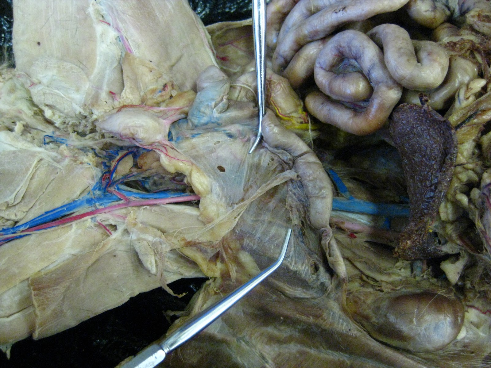
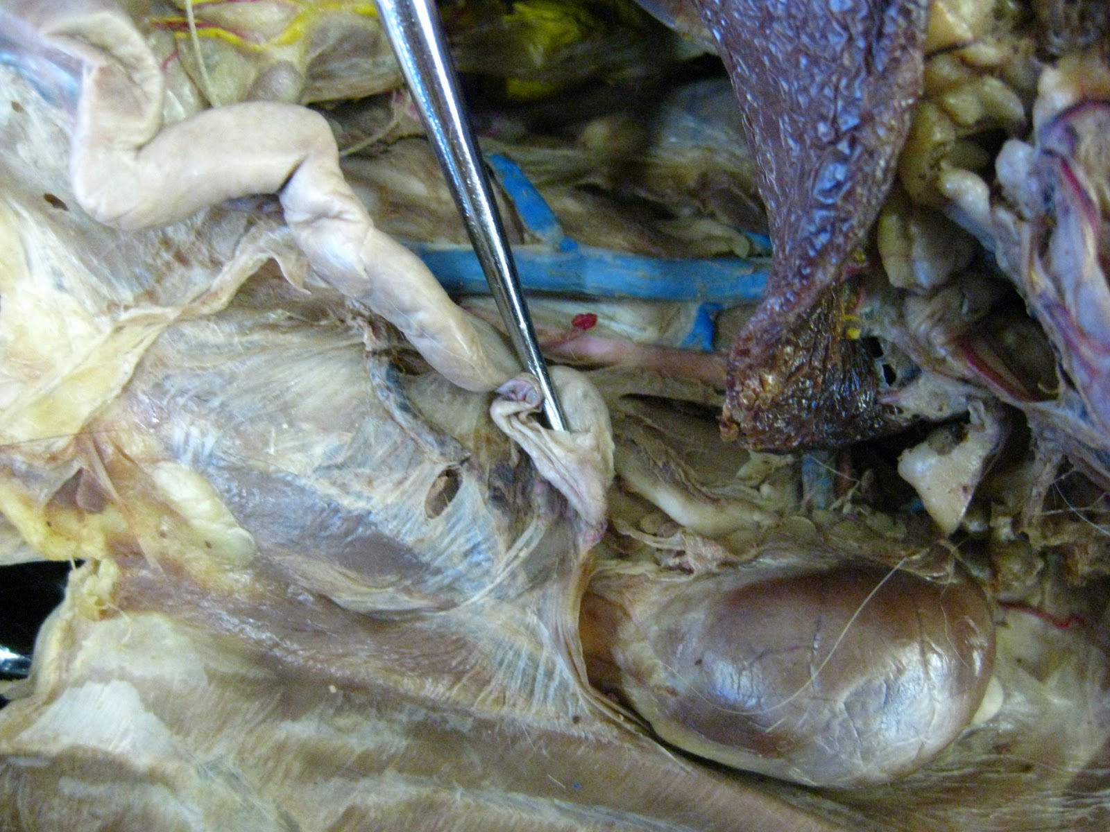
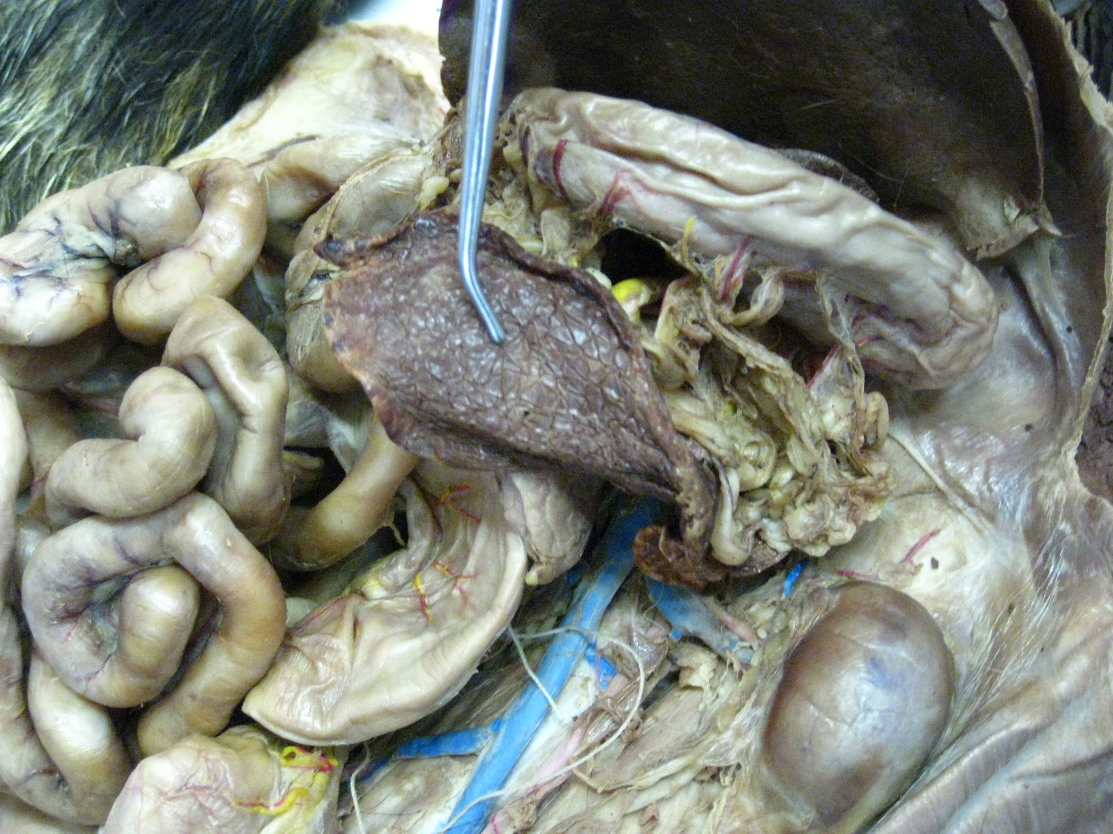
In the embalmed cat, the Spleen resembles dried fruit (burgundy color).

Sunday, November 27, 2011

Human Female Pelvis Model

Unlabeled Female Pelvis Model:


Female Model terms:

1. Uterus
2. Ovary
3. Oviduct (aka "Uterine tube" or "Fallopian tube")
4. Fimbriae
5. Vagina
6. Urinary Bladder
7. Urethra


Labeled Female Pelvis model:


Wednesday, November 9, 2011

Female Cat Reproductive System

uterus of female cat:
rough outline of the visible areas of the uterus of the female cat.


uterine body:

uterine horn:
Right and Left uterine horns indicated respectively by probes.

broad ligament (of cat uterus):
Broad ligament is not really a "ligament" (it is NOT made of Dense Regular CT and it does NOT link bone to bone); instead, broad ligament is made of Areolar connective tissue and Simple Squamous epithelium.

round ligament (of cat uterus):
Round ligament is not really a ligament; it is part of the Mesometrium (one of 3 distinct layers of the Broad ligament of the uterus).

uterine tube (aka "oviduct" , "Fallopian tube"):
The INFERIOR PROBE is pointing to the uterine tube coming off of the left infundibulum of the left ovary. The superior probe is resting on the left uterine horn.


infundibulum:
Infundibulum is the thin sheath covering the small "bean"-like ovary.


ovary:
Cat ovary looks like a small bean with a thin sheath over it (the infundibulum).


urethra (female cat):
Female cat's urethra begins directly inferior to the urinary bladder and ends where it anastomoses (joins) with the vagina, forming the urogenital sinus (unique structure to the female cat).


vagina:
Female cat's vagina is not entirely visible in this photo because it is deep to the urethra (superficial tube shown); instead, the space between the 2 probe ends designates where the vagina begins (immediately inferior to the uterus) and ends (at the beginning of the urogenital sinus, where the urethra and vagina first join together).


end of uterus (beginning of vagina):


 urogenital sinus:
Anastomosis of the urethra and vagina, forming a single, common vessel unique to the female cat.


Male Cat Reproductive System

penis of male cat (2 views):


scrotal sac (scrotum):

spermatic cord (both shown):


ductus deferens (aka "vas deferens"):

inguinal canal: no original lab picture (cat not sufficiently intact); opening/canal leading from the testis ("outside" of the body, held in the scrotal sac/scrotum) to the abdominal cavity. The inguinal canal contains the spermatic cord (conduit for blood vessels, lymphatic vessels, and ductus deferens of the testis). Link for inguinal canal picture from another site: inguinal canal of male cat.


prostate gland:
For the purpose of the Prof's Lab exam, the Prostate gland can simply be identified as the "bump" INSIDE the anastomosis (joining) of the male cat's urethra and its 2 ductus deferentes (plural of ductus deferens).


bulbourethral gland:
Bulbourethral gland secretes a portion of seminal fluid into the urethra and is, in general, located around the base of the penis; More accurately, the bulbourethral gland is really 2 glands--one is located on each side of and behind the male cat's urethra.

male cat's urethra:
Male cat's urethra is indicated by the probe immediately inferior to the prostate gland, which can be found inside the anastomosis of the male cat's urethra and 2 ductus deferentes.


tunica albuginea (of testis):
Tunica albuginea resembles a translucent/see-through "plastic wrap" over each testis.


epididymis ("ductus epididymis"):
Epididymis is the structure/duct immediately superior to the testis and directly connected to the ductus deferens (not visible here because it is encased by the spermatic cord "conduit").