Unlabeled Female Pelvis Model:

Female Model terms:
1. Uterus
2. Ovary
3. Oviduct (aka "Uterine tube" or "Fallopian tube")
4. Fimbriae
5. Vagina
6. Urinary Bladder
7. Urethra
Labeled Female Pelvis model:

 |
| rough outline of the visible areas of the uterus of the female cat. |

 |
| Right and Left uterine horns indicated respectively by probes. |
 |
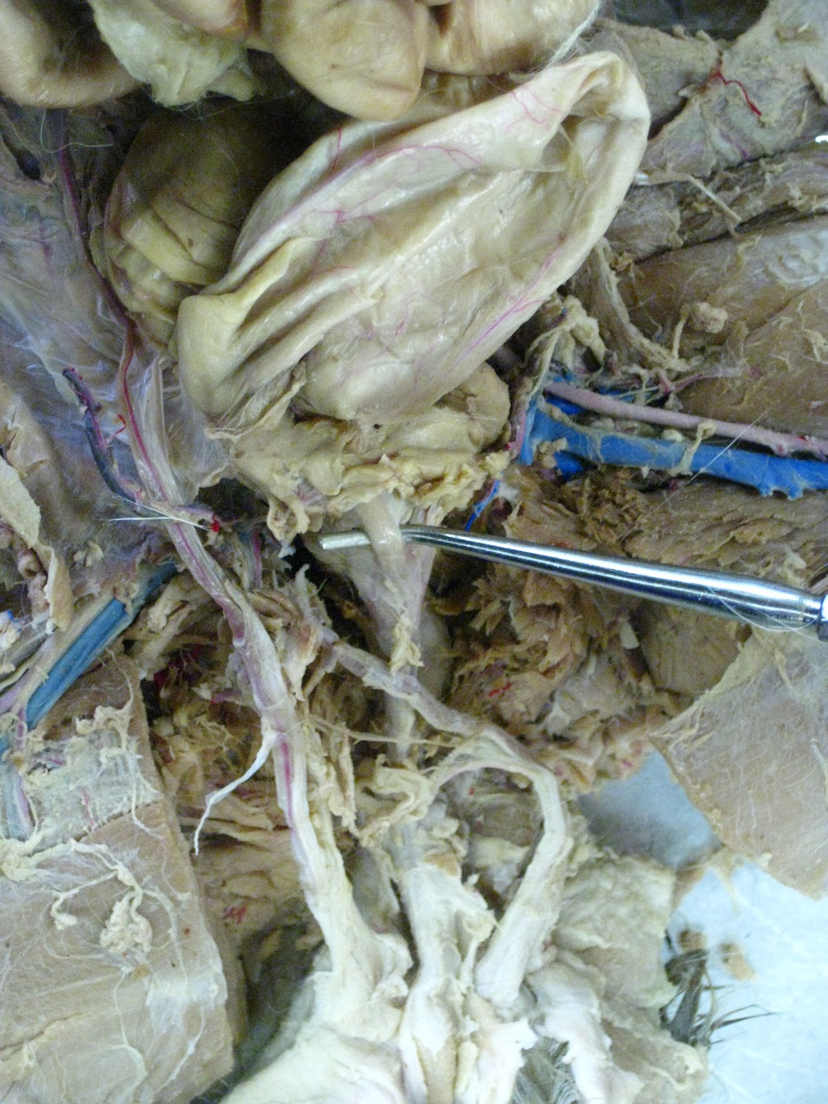
| Broad ligament is not really a "ligament" (it is NOT made of Dense Regular CT and it does NOT link bone to bone); instead, broad ligament is made of Areolar connective tissue and Simple Squamous epithelium. |
 |
| Round ligament is not really a ligament; it is part of the Mesometrium (one of 3 distinct layers of the Broad ligament of the uterus). |
 |
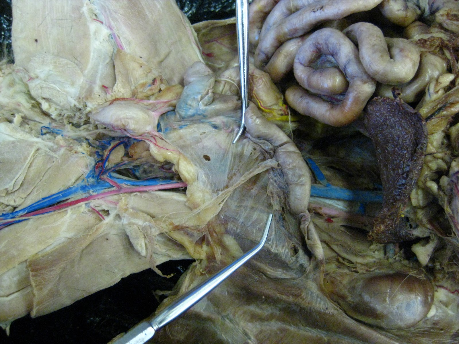
| The INFERIOR PROBE is pointing to the uterine tube coming off of the left infundibulum of the left ovary. The superior probe is resting on the left uterine horn. |
 |
| Infundibulum is the thin sheath covering the small "bean"-like ovary. |
 |
| Cat ovary looks like a small bean with a thin sheath over it (the infundibulum). |
 |
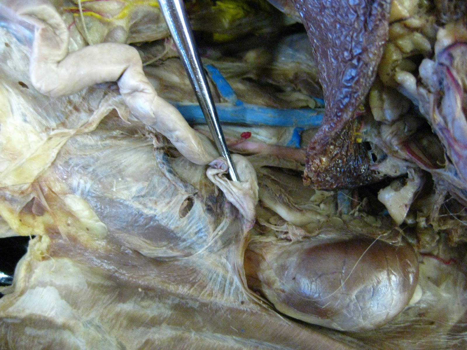
| Female cat's urethra begins directly inferior to the urinary bladder and ends where it anastomoses (joins) with the vagina, forming the urogenital sinus (unique structure to the female cat). |
 |
| Female cat's vagina is not entirely visible in this photo because it is deep to the urethra (superficial tube shown); instead, the space between the 2 probe ends designates where the vagina begins (immediately inferior to the uterus) and ends (at the beginning of the urogenital sinus, where the urethra and vagina first join together). |

 |
| Anastomosis of the urethra and vagina, forming a single, common vessel unique to the female cat. |


 |
| For the purpose of the Prof's Lab exam, the Prostate gland can simply be identified as the "bump" INSIDE the anastomosis (joining) of the male cat's urethra and its 2 ductus deferentes (plural of ductus deferens). |
 |
| Bulbourethral gland secretes a portion of seminal fluid into the urethra and is, in general, located around the base of the penis; More accurately, the bulbourethral gland is really 2 glands--one is located on each side of and behind the male cat's urethra. |
 |
| Male cat's urethra is indicated by the probe immediately inferior to the prostate gland, which can be found inside the anastomosis of the male cat's urethra and 2 ductus deferentes. |
 |
| Tunica albuginea resembles a translucent/see-through "plastic wrap" over each testis. |
 |
| Epididymis is the structure/duct immediately superior to the testis and directly connected to the ductus deferens (not visible here because it is encased by the spermatic cord "conduit"). |

 |
| adrenal gland is the roundish mass underneath the blue-stained vessel. |
 |
| renal arteries normally stain bright red, but did not receive a stain in this specimen. |
 |
| renal veins received a strong blue-stain. |
 |
| Urinary bladder of male cat. |
 |
| Urinary bladder of female cat. |


 |
| Male cat's urethra begins directly below (inferior to) the urinary bladder and ends where the penis ends. |
 |
| Female cat's urethra begins directly below (inferior to) the urinary bladder and then anastomoses (joins) with the vagina, forming the urogenital sinus (unique to the female cat). |
 |
| ductus deferens (right tube & left tube) where they anastomose with the urethra of the male cat, forming a thickened area: the prostate gland. |
 |
| ductus deferens before it reaches the anastomosis of the prostate gland. |

 |
| Urogenital sinus is formed by the anastomosis (joining of vessels) of the vagina and urethra of the female cat. |
 | ||||||||||||||||||||||||||||||||||||||||||||||||||||||||||||||||
| The prostate gland is located INSIDE the anastomosis of the 2 ductus deferens (plural = ductus deferentes) with the urethra of the male cat. |
 |
| area between the probes indicate the male cat's penis. |
 |
| head of penis of male cat. |


 |
| Items not labeled in photo: (1) Renal Hilum - indented area where ureter, renal artery, and renal vein enter the kidney. (2) Renal Capsule - outermost layer of model (burgundy color). (3) Renal Sinus - empty space inside the kidney that is occupied by the renal pelvis, renal calyces, blood vessels, nerves, and fat (adipose tissue). (4) dot-filled circle thing below the Renal Pelvis - it is simply another view of a Major Calyx (cross-sectioned cup). |